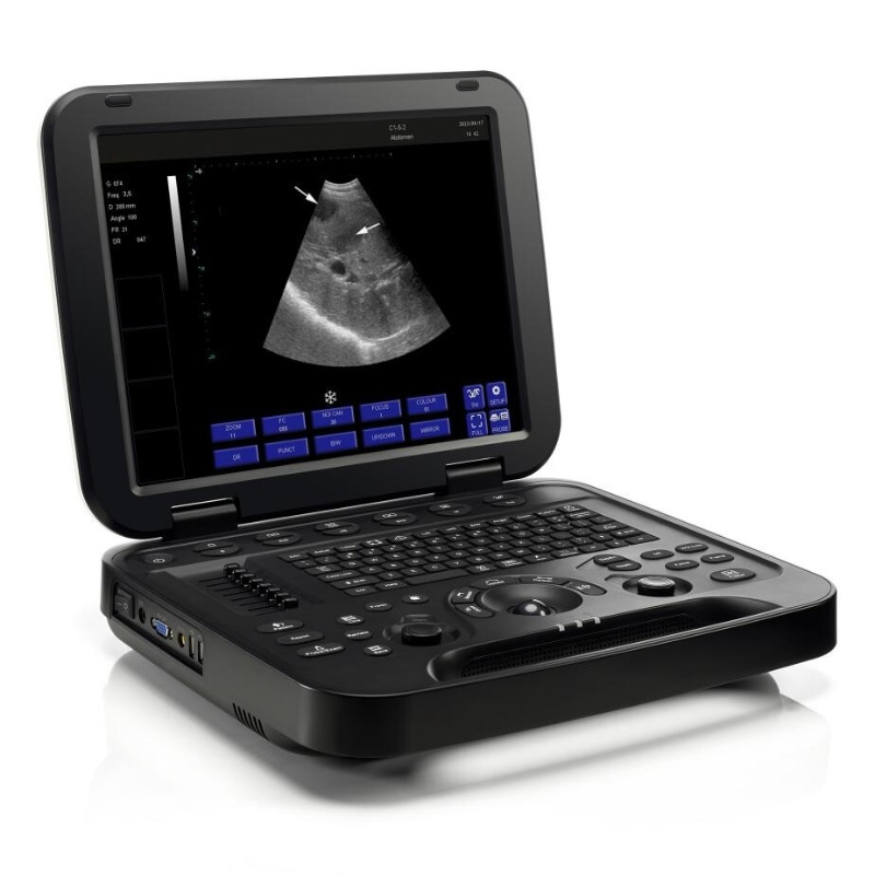
Product Display
Excellent performance for you
By combining advanced adaptive precise transmission and reception control with efficient multi-channel parallel processing technology, the machine’s computing speed is maximized while achieving the best image performance.
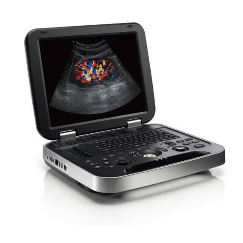
- Main Monitor 15 Inch HD LCD Display.
- Silicone backlit button design.
- Rich external interfaces.
- Portable design, light weight and small size.
- Dual transducer ports.
Product Features
- 1. Optimized Parameters Enhance Diagnostic Accuracy: 15″ high definition LCD, tilt
display, Quick Save One push image transfer to local or directly to, USB Stored images
can be edited and measured again, Picture-in-picture, partial zoom function, POptI™
Auto Optimization: single button image optimization, PStation™: clipboard, onboard
Reporting.
- 2. TSI Technology: TSI’s tissue-specific imaging function sets dedicated sound velocity
matching for different examination parts, such as muscles, liquids, fats, etc., to improve
the diagnostic value of obese animals or special diagnosis parts, making the ultrasound
image layer clearer and the particles finer. , the resolution is better.
- PrismX™Spatial Composite Imaging:By employing PrismX™ spatial imaging technology, our system enhances contrast, fine resolution, and spatial resolution.It optimizes tissue and pathological interface echo continuity while minimizing artifacts such
as specular reflection, speckle, scattering, and attenuation, ensuring superior image
clarity.
- PanoramaVision™ Extended Field Imaging: Panorama Vision™ technology
captures a series of two dimensional sectional images, which are reconstructed into an
ultra- wide sectional image with a continuous field of view. This enables precise measurement of organ size and lesions, while providing a comprehensive view of the lesion’ s scope, internal echo, location, size, and adjacent tissues.
- 5. PureSight™ Imaging Technology: Our PureSight™ technology reduces speckle
artifacts,reduces speckle artifacts,improves smoothness along preferred edge directions, and maintains consistent local average gray levels. This results in
clearer and more visually appealing images, enhancing diagnostic confidence.
- 6. Anatomic M-mode echocardiography: Anatomic M-type echocardiography can also
be used to diagnose complex heart diseases, such as congenital heart defects, valvular heart disease, etc. Through detailed observation and analysis of the heart structure,
doctors can more accurately judge the type and extent of the disease and develop a more appropriate treatment plan for animals.
- 7. Full intelligent puncture enhancement:The intelligent blue-light enhancement
technology automatically detects the needle, optimizing image quality for safer, precise,
and efficient puncturing.
- 8. Specialized musculoskeletal examination mode:This new multi-beam configuration
offers a professional MSK inspection mode with pre-optimized conditions for clearer, intricate images, reducing adjustments and boosting efficiency.
- 9. Powerful features and measurement package: Using advanced digital
beamforming¶llel signal processing, we offer tissue harmonic, ladder scopic, wide
field, elastic imaging, professional measurement tools for an integrated MSK solution.
Technical Specifications
|
PARAMETER
|
SPECIFICATION
|
|
Application: Generic, Abdomen, Obstetrics, Gynecology, Vascular, Small Parts, Urology, MSK, Cardiology.
|
|
Transducer Types
|
Convex,Linear, Micro convex probe.
|
|
Operating Modes
|
B Mode ,Dual B, Quad B, CF Mode, M Mode B/M Mode,PW Mode Power Doppler Imaging, Directional Power Doppler Imaging
Wide View (Extend Field of View)(Optional),Picture in Picture Trapezoid, Panorama, Color M Mode, Anatomy M Mode
Needle Enhance Mode(Optional), Contrast Enhance Mode(Optional)
|
|
Imaging Technique & Function
|
Automatic optimization
Spatial compound imaging
Speckle reduction imaging
Color quantification
Fine angle steer
Tissue harmonic imaging (THI), pulse inversion harmonic imaging (iTHI)
Virtual convex
Animal information database
Image archive on integrated hard drive
Real-time automatic Doppler calculation
OB calculations
Fetal trending
Multi-gestational calculations
Hip dysplasia calculations
Vascular calculations
Urological calculations
Renal calculations
On-board electronic documentation
PW Auto-trace
Privacy and security
DICOM
Wide view
External USB printer connection
|
|
Simultaneous Capability
|
B/CF, B/PDI or DPDI, B/PW,C/M, B/M,B+CF or PDI or DPDI/PW or M.
|
|
Monitor
|
15 inch High Resolution LCD Monitor Resolution: 1024*768
Silicone button design
Magnetic latch closure
Built-in speaker
Brightness and Contrast adjustable
|
|
Convex probe
|
Applications:
Generic, abdomen,, urology
|
|
Frequencies:
3.5:2.5–6M
|
|
Linear probe
|
Applications:
Small parts, vascular,musculoskeletal, superficial
|
|
Frequencies:
L40: 5–12M
|
|
Microconvex probe
|
Applications:
Generic, abdomen,, urology
|
|
Frequencies:
5.0:5–9M
|
Product Configuration
1.Standard Configuration:
Power cable, Operator’s manual.


 Contact
 Add to List