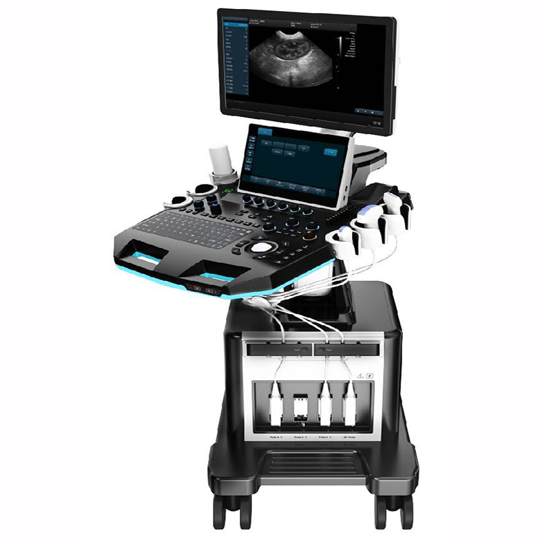
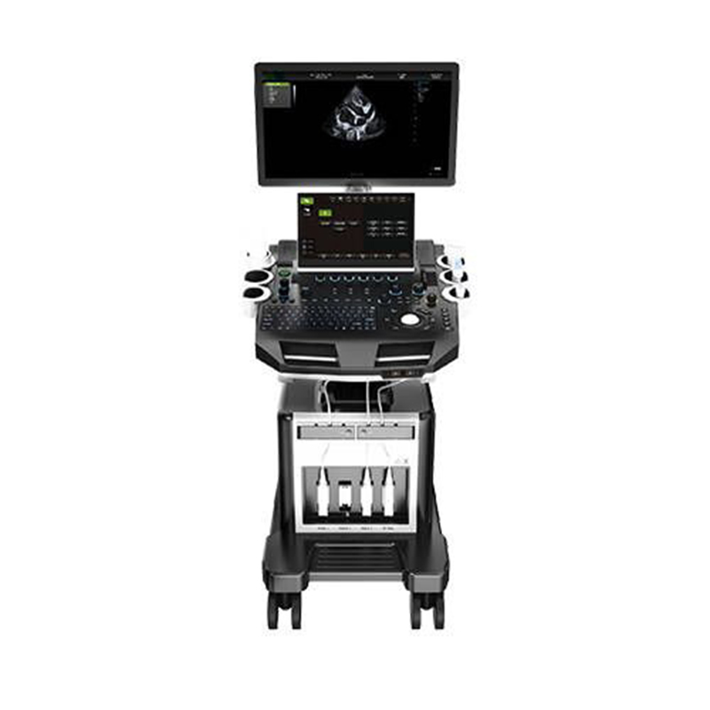
Product Display
Excellent performance for you
By combining advanced adaptive precise transmission and reception control with efficient multi- channel parallel processing technology, the machine’s computing speed is maximized while achieving the best image performance.
-23 inches high resolution LCD Monitor
-Imaging Technique:
Automatic optimization;
Spatial compound imaging;
Speckle reduction imaging;
Color quantification;
Fine angle steer;
Tissue harmonic imaging;
Pulse inversion;
Harmonic imaging
-One-key optimization to optimize image in real time.
Auto trace, measurement and calculation Large hard drive with RAM8G, ROM256G support large cases and long-term storage.
-Triplex: B+CF/PDI/DPDI+PW
-Support multi-species obstetric biometric calculation (bovine, equine, canine, feline, ovine, porcine)
-Large hard drive with RAM8G, ROM256G support large cases and long-term storage.
Product Features
Blood vessels; Nerves; Clinical applications in abdomen; Heart; Urinary tract;
Musculoskeletal system
Function
Real-time automatic Doppler calculation, OB calculations, Multi-gestational calculations, Hip dysplasia calculations,Vascular calculations, Urological calculations, Renal calculations, Cardiac calculations, On-board electronic documentation, PW Auto-trace, Privacy and security, DICOM, Wide view, Auto IMT.
Cine/Image
Scrolling timeline memory, Thumbnails Image/Cine display, Cine review loop, Cine review speed, On-board database of patient information from past exams.
Optional Display Mode
Tissue Doppler Imaging, Wide View, Needle Enhance Mode.
Standard Display Modes
B Mode, Dual B, Quad B, CF Mode, M Mode, B/M Mode, PW Mode, Power Doppler Imaging, Directional Power Doppler Imaging, Picture in Picture, Trapezoid, Panorama, Color M Mode, Anatomy M Mode.
Built-in case management system
Which can be viewed according to different labels; one-click editing of report templates, and support for direct connection to various types of printers.
Tissue Doppler Imaging
Using the principle of Doppler effect to show the movement of myocardial tissue.
Technical Specifications
|
PARAMETER
|
SPECIFICATION
|
|
Application: General, Abdomen, Obstetrics, Reproduction, Vascular, Small Parts, Urology, MSK, Cardiology.
|
|
Transducer Types
|
Convex,Linear, Micro convex, Phased array probe
|
|
Transducer Types
|
B Mode ,Dual B, Quad B, CF Mode, M Mode, B/M Mode,PW Mode
Power Doppler Imaging,Directional Power Doppler Imaging, Picture in Picture, Trapezoid, Panorama, Color M Mode, Anatomy M Mode
|
|
Imaging Technique
|
Automatic optimization
Spatial compound imaging
Speckle reduction imaging
Color quantification
Fine angle steer
Tissue harmonic imaging
Pulse inversion harmonic imaging Virtual convex
|
|
Image formats
|
AVI, BMP, JPG, PNG, DICOM
|
|
System Language Support
|
English, Arabic, Chinese. French. German,Indonesian, Italian, Portuguese, Russian, Spanish
|
|
Triplex
|
B+CF, PDI, DPDI+PW
|
|
Convex probe
|
Applications:
Generic, abdomen, urology
|
|
Frequencies:
3.5:2.5–6M
|
|
Linear probe
|
Applications:
Small parts, vascular,musculoskeletal, superficial
|
|
Frequencies:
L40: 5–12M
|
|
Microconvex probe
|
Applications:
Generic, abdomen, urology
|
|
Frequencies:
5.0:5–9M
|
|
Phased array probe
|
Applications:
Generic, abdomen, cardiology
|
|
Frequencies:
5.0:3–7M
|
Advanced Imaging Technologies
COMPOUND:Image optimization adapt to different applications
iTHI:Contrast, resolution & SNR improvement
SRI:Speckle reduction & Smooth the tissue images


 Contact
 Add to List